Community Acquired Pneumonia: When to Admit and How to Treat

In the unit Respiratory Tract Infections: Pneumonia we covered the assessment of a patient presenting with signs and symptoms of pneumonia including differentiating between the 3 main types. In the unit Community Acquired Pneumonia: Outpatient Treatment we talked about those patients who are stable enough to be treated at home. This unit focuses on the treatment of patients with community acquired pneumonia who have a risk of morbidity or mortality great enough to warrant treatment in the hospital.

We can objectively assess where a patient with community acquired pneumonia should be treated based on the CURB 65 score.

CURB 65

The preferred way to asses whether hospital admission is required for pneumonia is the Pneumonia Severity Index (PSI). PSI estimates the mortality of adult patients with CAP. It is complicated to calculate. It requires alot of patient history and labs that are not readily available in the outpatient setting including pH and partial pressure of oxygen.

Instead the CURB 65 score is used to assess risk of mortality and therefore the level of care (inpatient, outpatient) in patients presenting with CAP.

Illustration of the components of the CURB 65 score to assess the risk of mortality in patients with community acquired pneumonia

CURB-65 assigns 1 point for each of the 4 assessment points.

The urea level is the blood urea nitrogen (BUN). Normal levels are 5-20mg/dL. Because it requires a lab draw, BUN is not always readily available in many outpatient settings. There is an abbreviated score called CRB-65 that eliminates the urea component.

Respiratory rate >30 breaths per minute is 1 point.

Blood pressure <90/60 mmHg is 1 point

Age > 65 is 1 point

A CURB-65 score of 2 or more s a high risk for morbidity and/or mortality therefore the need for hospital admission.

MedCalc offers a CURB 65 score calculator.

At this point we’ve answered 2 of the 3 “where” questions of pneumonia.

  1. Where the bug was contracted: community or within 48 hours of hospital admission
  2. Where the patient will be treated: hospital (inpatient, CRB>2)

At this point we need to decide another where. Where in the hospital will they be treated. Whether they go to ICU or general medicine floor requires assessment of the severe cap criteria.

Decision tree for where a patient with community acquired pneumonia should be treated
Severe CAP Criteria

This is a clinical tool designed to predict the probability of ICU admission. It includes criteria that can be readily assessed in any emergency room. The CAP criteria is divided into 2 categories: major and minor.

Criteria for patients who should be treated in the ICU

The major criteria are straightforward. If you patient is hypotensive enough that they require the use of vasopressors or requires mechanical ventilation due to respiratory failure, the recommendation is that your patient is admitted directly to intensive care.

If your patient requires hospitalization (CURB/CRB>2) but does not require vasopressors or mechanical ventilation, we assess minor criteria.

Minor Criteria for patients who should be treated in ICU or on general medical floor

The ATS/IDSA guidelines define severe community acquired pneumonia as either one major criteria or atleast 3 minor criteria. 3 or more minor criteria suggest the need for higher levels of care like ICU or telemetry. Everyone else can admitted to general medical floor if there are no confounding complications. Always use clinical judgement in conjunction with these prognostic tools to determine the level of care.

Just like the decision to treat inpatient versus outpatient, there are cost considerations and procedural risks associated with ICU stays. On the other hand, there is a higher rate of mortality when a patient is transferred from the floors to the units versus direct admission to the units.

Assess History of Infection

We now know where in the hospital our patient will be treated. Before we select antibiotics for inpatient treatment we have to assess their history of infection. Whether you’ve decided ICU or general medicine, all adults treated inpatient must be have they history checked for:

History of exposure that should be assessed for all patients with pneumonia
  • history of respiratory MRSA in the last 12 months
  • history of pseudomonas in the past 12 months
  • history of hospitalization with IV antibiotics in the last 3 months

If your patient has none of these they are considered as having no significant history with regards to pneumonia.

These 4 criteria will determine what antibiotics will be used. Like we did for outpatient treatment of CAP we will look at clinical scenarios and treatments regimens for each sub category of inpatient CAP.

Refer to Respiratory Tract Infections: Pneumonia to review what bacteria we are targeting in each type of pneumonia.

General Medical: Inpatient

Clinical Scenario 1:

Your patient has no history of MRSA or pseudomonas in the last year and no hospital admission with IV antibiotics in the last 90 days.

treatment algorithm for treatment of community acquired pneumonia with no risk factors for multidrug resistant bacteria
Clinical Scenario 2:

Your patient has a history of respiratory MRSA in the last 12 months.

treatment algorithm for treatment of CAP with risk factors for MRSA
Clinical Scenario 3:

Your patient has a history of respiratory pseudomonas in the last 12 months

treatment algorithm for treatment of community acquired pneumonia with no risk factors for multidrug resistant bacteria pseudomonas

These regimens above will provide the coverage of the regimen in scenario 1 + pseudomonas coverage.

Clinical Scenario 4:

Your patient has a history of respiratory MRSA and pseudomonas in the last 12 months

treatment algorithm for treatment of community acquired pneumonia with no risk factors for multidrug resistant bacteria
Clinical Scenario 5:

Your patient has a history of hospitalization with intravenous antibiotics in the last 90 days

  1. Use standard regimen (clinical scenario 1)
  2. Get sputum gram stain and culture
  3. Escalate to coverage for MRSA or pseudomonas based on culture/gram stain results

Clinical scenario 5 will gain more context as we move on to community acquired pneumonia patients who require higher levels of care than general medicine in the next unit.

Alternative Antibiotics

When are they needed?

Considerations for choice of antibiotics: coverage, allergy, cost utility and formulary restriction

The primary regimen provided in each clinical scenario include antibiotics that are fairly common throughout the United States. A common reason you will have to deviate from this standard regimen is allergy. Pneumonia guidelines are laced with beta lactams.

Unfortunately penicillin is also the most commonly reported drug allergy. Whether these are true anaphylactic responses or simply drug intolerances/unfavorable side effects is another discussion in itself.

Some hospitals preferentially use antibiotics that have lower dosing frequencies or requires less management. Azithromycin, for example, is dosed daily, clarithromycin is dosed twice a day. Vancomycin requires patient specific dosing and fairly frequent monitoring of labs and levels. Linezolid does not.

Certain antibiotics may be restricted to use only by infectious disease doctors due to their cost and spectrum of coverage. Those antibiotics are usually referred to as the “big guns” and are reserved for especially resistant strains of bacteria like extended spectrum beta lactamase resistance or Acinetobacter.

Know what antibiotics are on your hospitals formulary and which have restricted access. Be familiar with the alternative antibiotics listed below the primary regimen. It is useful to know what antibiotic you would recommend in the event of a penicillin allergy.

Sputum Cultures:

When are they needed in CAP?

The ATS/IDSA state that they are “neither for or against routinely obtain sputum gram stain and cultures in all adult patients treated in a hospital setting.”

They however specifically recommend sputum gram stain and cultures in severe CAP (ICU admission) and when there are risk factors for MRSA and pseudomonas (history for prior infection in the last 12 months or history intravenous antibiotics in the last 3 months).

If there is an actual documented history of MRSA and/or pseudomonas: start empiric antibiotic coverage and deescalate if the sputum results are negative.

If they have no history of respiratory MDR bacteria but are at risk for MRSA and/or pseudomonas due to exposure then wait for culture results before extending coverage.

Gram stains usually result in 24 hours, culture results in 24-48 hours.

Blood Cultures:

When are they needed in CAP?

ATS/IDSA guidelines recommended that blood cultures be drawn PRIOR to initiation of antibiotics only in those same patient we just discussed for sputum cultures. These are Hospitalized patients with:

  1. severe CAP (ICU admission)
  2. history of respiratory MRSA and/or pseudomonas in the past 12 months
  3. recent exposure to MRSA and/or pseudomonas exposure i.e. hospitalization with intravenous antibiotics in the past 3 months

If we’re covering MRSA and/ or pseudomonas for treatment of CAP we need blood and sputum cultures.

Duration of Treatment

The ATS/IDSA guidelines are pretty straightforward with regard to the duration of treatment for community acquired pneumonia.

  • Treatment should be no less than 5 days even if patient is clinically stable before then
  • If CAP is due to MRSA or pseudomonas then treat for 7 days

This study unit is a hefty one. Hopefully the way the guidelines have been deconstructed here will make it easier to apply in your practice. CAP is one of those disease states you will see over and over again. Use the aids and illustrations as a reference and soon it will become second nature.

Subscribe

Subscribe to get the latest study unit in your inbox.

The information on this website is intended to be used solely for educational and informational purposes. While the content may be about specific medical and health care issues, it is not a substitute for or replacement of personalized medical advice and is not intended to be used as the sole basis for making individualized medical or health-related decisions.

How to Manage Pneumonia in the Outpatient

Pneumonia is simply a type of infection of one or both lungs.

Community acquired pneumonia (CAP) describes the clinical scenario where signs and symptoms of a lung infection started outside of the hospital OR within 48 hours (2 days) of hospital admission.

Within this timeframe, it is likely the patient encountered the bug and it has been incubating prior to being admitted.

The first part of this series, Respiratory Tract Infections: Pneumonia covers the basics of pneumonia. It explains the different types of pneumonia, how we assess a patient and the 3 “where” questions that must be answered when a patient presents with sign and symptoms of pneumonia. It sets the foundation for understanding how we treat each subset of pneumonia.

The following chart provides a general summary of the content covered in that unit. Details on all the steps illustrated can be found here.

Decision tree for where a patient with pneumonia should be treated: inpatient versus outpatient

For this study unit we will assume that our patient had signs and symptoms of pneumonia outside of the hospital or within 48 hours of hospital admission: the diagnosis is community acquired pneumonia (CAP).

Let us first consider those patients with a CURB-65 score of <2 i.e. patients suitable for treatment of pneumonia in outpatient settings.

Treatment of CAP: Outpatient

We have a diagnosis (pneumonia) based on sign/symptoms, labs and imaging. We know where the infection started (outside of hospital/community) and where the patient will be treated (outpatient, CURB/CRB<2).

At this point there are only 2 clinical scenarios for treatment that is dependent on whether our patient has disease states that place them at risk of infection by resistant bugs. Theses includes:

How to assess CURB 65 to determine if a patient needs to be treated inpatient versus outpatient
Clinical Scenario 1: CAP Outpatient

Your patient has NO disease states that places them at risk for potential infection by resistant bugs.

Treatment algorithm for outpatient treatment of CAP no risk factors

In otherwise healthy patients the guidelines recommend treatment of pneumonia in outpatient with amoxicillin. If patient has an allergy we can use doxycycline, azithromycin or clarithromycin.

Clinical Scenario 2: CAP Outpatient

Your patient has one or more of these diseases that places them at risk for potential infection by resistant bugs.

Treatment algorithm for outpatient treatment of CAP with risk factors

If your patient has one or more of the comorbidities listed, then we can use either combination therapy or monotherapy to treatment pneumonia in outpatient. In the illustration above you can substitute any of the drugs in the primary regimens for an alternative listed in the corresponding category.

Please note that there are some cases where a patient has a CURB/CRB score of <2 and requires hospitalization. For instance, a patient may not be able to tolerate oral medications. They may have a lifestyle or home environment that has a high likelihood of not reliably completely antibiotics like drug abuse, cognitive impairment or functional impairment. Clinical judgement must always be used in conjunction with validated prognostic tools.

Alternative Antibiotics

When are they needed?

The primary regimen provided in each clinical scenario includes antibiotics that are fairly common throughout the United States. A common reason you will have to deviate from this standard regimen is allergy.

Pneumonia guidelines are laced with beta lactams. Unfortunately penicillin is also the most commonly reported drug allergy. Whether these are true anaphylactic responses or simply drug intolerances/unfavorable side effects is another discussion in itself.

It is a good idea to know a non-penicillin antibiotic for each clinical scenario so that you are prepared when the the first line agent is not an option. Sometimes, in the inpatient setting, a beta lactam may be given despite a listed penicillin allergy with close monitoring. This is not an option in the outpatient setting because your patient will be taking the medication at home.

Sputum Cultures:

When are they needed in CAP?

Sputum cultures are NOT recommended for CAP managed in the outpatient setting. Most clinics will not have the staff needed to obtain sputum cultures. As we’ve discussed earlier, the like causative agents in CAP are going to be susceptible because there is relatively less antibiotic exposure to foster conversion to MDR bugs.

Guidelines consider the cost utility of the interventions that are recommended. Getting cultures on samples that will routinely be susceptible adds unnecessary costs to the outpatient treatment of pneumonia.

Blood Cultures:

When are they needed in CAP?

ATS/IDSA guidelines recommended that blood cultures be drawn only in patients hospitalized with CAP. We will discuss patients with CAP requiring hospitalization in a different study unit.

Duration of Treatment

The ATS/IDSA guidelines are pretty straightforward with regard to the duration of treatment for CAP

Treatment should be no less than 5 days even if patient is clinically stable before then i.e. complete the course of therapy, even if patient starts to feel better before it ends

This unit serves a good launching pad to dive into the diagnosis and treatment guidelines for community acquired pneumonia from the American Thoracic Society (ATS) and IDSA.

Hopefully the way the guidelines have been deconstructed here will make it easier to apply in your practice. CAP is one of those disease states you will see over and over again. Use theses aids and illustrations as a reference and soon it will become second nature.

Subscribe

Subscribe to get the latest study unit in your inbox.

The information on this website is intended to be used solely for educational and informational purposes. While the content may be about specific medical and health care issues, it is not a substitute for or replacement of personalized medical advice and is not intended to be used as the sole basis for making individualized medical or health-related decisions.

Navigating Pneumonia: Where and How to Treat

Pneumonia is an infection of the lower respiratory tract specifically the lungs. You can refer to the introduction of the study unit on SINUSITIS for a detailed discussion of the respiratory system.

This study unit is based on the current Clinical Practice Guideline of the American Thoracic Society and Infectious Diseases Society of America (ATS/IDSA) for the treatment of community acquired pneumonia (CAP), hospital acquired pneumonia (HAP) and ventilator associated pneumonia (VAP).

Cross section of the torso and head differentiating the upper respiratory tract from the lower respiratory tract

Infectious organisms can enter the lower respiratory tract in 1 of 3 ways:

  1. direct inhalation
  2. aspiration from the throat (oropharyngeal)
  3. via the blood from another site of infection (hematogenous transfer)

Community Acquired Pneumonia

Community acquired pneumonia (CAP) describes the clinical scenario where signs and symptoms of a lung infection started outside of the hospital OR within 48 hours (2 days) of hospital admission. Within this timeframe, it is likely the patient encountered the bug and it has been incubating prior to being admitted.

Causative Organisms

The most common cause of community acquired pneumonia is a viral infection. Rhinovirus and influenza are the 2 most common viral causes. As a reminder, antibiotics have no effect on viruses. Unnecessary treatment with antibiotics will only increase the potential for developing multidrug resistant bacteria that are hard to treat if they become pathogenic.

Bacterial pathogens often coexist with viruses. There is no current test that can quickly and accurately distinguish viral CAP from bacterial CAP. In the study unit covering community acquired pneumonia, we will talk about “double sickening” where an infection starts off as viral and then transitions to bacterial. The ATS/IDSA guidelines recommend treating empirically for possible bacterial infection or coinfection.

Chat showing the likely causative bacterial and viral agents of community acquired pneumonia

The most common bacterial pathogen in community acquired pneumonia is Streptococcus pneumoniae.

Haemophilus influenzae, Mycoplasma pneumoniae, Staphylococcus aureus, Legionella species, Chlamydia pneumoniae, and Moraxella catarrhalis are the other common bacterial agents.

Hospital Acquired Pneumonia

Hospital acquired pneumonia (HAP) describes the clinical scenario where signs and symptoms of a lung infection started at least 48 hours (2 days) after hospital admission. Within this timeframe, it is likely that the patient contracted the bug from a source in the hospital.

Ventilator Acquired Pneumonia

Ventilator associated pneumonia (VAP) describes the clinical scenario where signs and symptoms of a lung infection started after at least 48 hours after intubation.

Causative Organisms

The common bacterial pathogens are the same for HAP and VAP. They vary by institution and is influenced by antibiotic use practices at the particular site. These bugs include  Pseudomonas aeruginosa,  Escherichia coli,  Klebsiella pneumoniae,  Enterobacter,  Acinetobacter, Staphylococcus aureus and Streptococcus.

Chart showing the likely causative agents in hospital acquired pneumonia.

Why does “WHERE” matter so much?

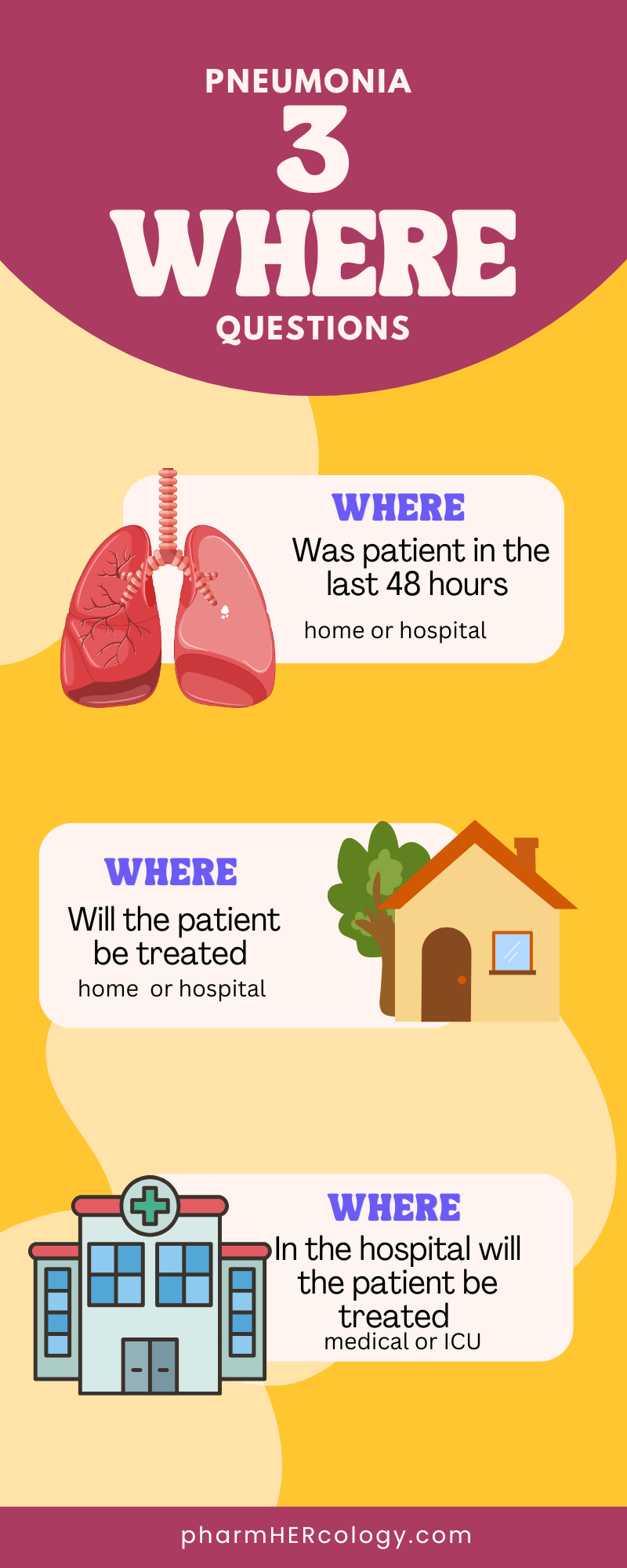
The antibiotic we use for the treatment of pneumonia is dependent on 3 “where” questions: where the bug was contracted and where the patient will be treated (home versus hospital) and where in the hospital they will be treated.

Infographic showing the 3 questions that need to be answered in all patients who present with signs and symptoms of pneumonia. Location in the last 48 hours, whether they will be treated at home or need hospital care. If they need hospital care, do they need general medical care or intensive care.

The setting where infection occurred is used to categorize pneumonia and the associated distinction between CAP/HAP/VAP is an important one. As we will see, it informs the choice of antibiotics and duration of therapy. This is because where it was contracted gives us a best-guess of the likely causative agent. Specifically whether we need to cover potentially resistant bacteria.

Where the patient will be treated (outpatient versus inpatient) will also determine the route of administration.

Watch on YouTube

Classification of Pneumonia

Chart illustrating the 3 classifications of pneumonia: community acquired, hospital acquired, or ventilator associated.

The clinical presentation of HAP and CAP are fairly similar. This creates the potential to overtreat CAP and breed resistant bugs or undertreat HAP and increase risk of mortality. This is why “WHERE” infection occurred is the first important question you must answer in the treatment of pneumonia.

Multi Drug Resistant Bacteria

Because there is greater, more consistent use of antibiotics within a hospital, there is greater potential for the development of multi drug resistant (MDR) bacteria. These organism like methicillin resistant staphylococcus aureus (MRSA) and pseudomonas aeruginosa (pseudomonas) are associated with higher mortality and require targeted treatment.

Illustration of how antibiotic resistance develops.

We do not want to expose patients who are unlikely to be infected with MDR bacteria with antibiotics used for these bugs. Whenever pathogens are exposed to antibiotics, over time they develop genetic mutations. These mutations allow that bug to circumvent the effect of the drug, thus developing resistance. This process is accelerated as more antibiotics are used in more people.

HAP/VAP and CAP have alot in common in terms of presentation and diagnosis but they will be addressed in different study units. They are distinct infections that have different therapeutic considerations.

Diagnosing Pneumonia

A diagnosis of pneumonia is based on clinical presentation and radiographic imaging (chest X ray).

How does a patient with pneumonia present?
Illustration of the 4 categories we would use to diagnose pneumonia: sign and symptoms. physical exam, labs and imaging.

Signs and symptoms refer to what the patient complains of. For pneumonia this includes fever, chills, shortness of breath, a productive cough and/ or blood in sputum.

The physical exam may show shortness of breath (tachypnea), increased heart rate (tachycardia), diminished breath sounds and/ or crackling when they take a breath (inspiratory crackles).

Their labs can show elevated white blood cells (leukocytosis) which is a classic sign of infection.

Once pneumonia is suspected based on patient presentation the guidelines recommend confirmation of pneumonia via a chest X Ray. Be aware that in most cases imaging does not occur in the outpatient setting for the diagnosis of pneumonia. Most clinics do not have onsite radiography.

The chest X ray will show infiltrates in the lungs.

When we see infiltration in the lungs it means there is something that is more dense than air like pus or blood.

Infiltration with signs and symptoms of infection, as explained above, suggests pneumonia.

The ATS/IDSA guidelines recommend a chest X ray for all adult patients with suspected pneumonia.

A chest x ray showing infiltration which is indicative of pneumonia.

Using patient’s sign and symptoms, physical exam, labs and chest X ray we can make a diagnosis of pneumonia.

At this point we know what to treat. Before we can select the appropriate antibiotics we need to answer the second “where”: where to treat. To objectively determine whether a patient would be best treated inpatient or outpatient we clinical prediction tools: CURB 65 and the (Pneumonia Severity Index) PSI scale.

Where to Treat Pneumonia

At this point we know where our patient encountered in the infecting organism. The next step is to determine where our patient needs to be treated: outpatient, inpatient or inpatient ICU.

CAP or HAP is not an indication of disease severity or where they get treated.

A patient with either one of these has the potential to be treated outpatient, inpatient or in critical care.

Where they are treated is based on clinical presentation and risk of mortality.

A patient with VAP will of course be treated in the hospital because they are on a ventilator. A patient with HAP may develop signs and symptoms after recent discharge from the hospital. Whether they need some be treated outpatient or inpatient depends on their clinical presentation (difficulty breathing, low oxygenation, organ failure).

Unnecessary hospital admission for cases that could be treated outpatient results in high costs for both patients and hospitals. It can also place the patient at risk for complications associated with hospital admission including exposure to MDR bacteria, and increased risk if blood clots.

The implications of not admitting a patient who needs additional support is more straight forward, it increases the risk of morbidity and mortality.

The Problem with PSI

The preferred way to asses whether hospital admission is required for pneumonia is the Pneumonia Severity Index (PSI). PSI estimates the mortality of adult patients with CAP. It is complicated to calculate. It requires alot of patient history and labs that are not readily available in the outpatient setting including pH and partial pressure of oxygen.

CURB 65

Instead the CURB 65 score is used to assess risk of mortality and therefore the level of care (inpatient, outpatient) in patients presenting with CAP.

Illustration of the components of CURB 65. Confusion, urea, respiratory rate and blood pressure.

CURB-65 assigns 1 point for each of the 4 assessment points.

The urea level is the blood urea nitrogen (BUN). Normal levels are 5-20mg/dL. Because it requires a lab draw, BUN is not always readily available in many outpatient settings. There is an abbreviated score called CRB-65 that eliminates the urea component.

Respiratory rate >30 breaths per minute is 1 point.

Blood pressure <90/60 mmHg is 1 point

Age > 65 is 1 point

A CURB-65 score of 2 or more suggests a high risk for morbidity and/or mortality therefore the need for hospital admission.

Decision tree for where a patient with pneumonia will be treated: inpatient versus outpatient.

Once we’ve made the decision to treat inpatient we need to decide where in the hospital to treat. Is the patient stable enough for the medical floor or does the patient need the additional support of the intensive care unit. This too will determine the choice of antibiotics. We will delve into this in the individual study units.

After answering those “WHERE” questions we can begin appropriate treatment.

The study units for CAP and HAP/VAP will review the specifics for these disease states including appropriate antibiotics, need for cultures, duration of treatment and much more.

I hope this study unit has set a strong foundation for the assessment of pneumonia. Pneumonia is one of those disease states you will see over and over again. Refer to the diagrams presented here often and it will soon become second nature.

Subscribe

Subscribe to get the latest study unit in your inbox.

The information on this website is intended to be used solely for educational and informational purposes. While the content may be about specific medical and health care issues, it is not a substitute for or replacement of personalized medical advice and is not intended to be used as the sole basis for making individualized medical or health-related decisions.

Sinusitis: What You Need to Know to Maximize Patient Outcomes

A respiratory tract infection is an infection that affects any part of the respiratory system. Sinusitis in an infection of the sinuses.

The parts of the respiratory system can be categorized as part of the upper respiratory tract or the lower respiratory tract.

Illustration of the 6 components of the respiratory system

The upper respiratory tract includes the:

  1. the sinuses
  2. pharynx (throat)
  3. larynx (voice box)
  4. nasal cavity

The lower respiratory tract includes:

  1. trachea (wind pipe)
  2. lungs

The Sinuses

The sinuses are air filled cavities that are located on either side of the nose and above the eyebrows. They are lined with cells that produce mucus. This mucus drains from the sinus cavities and down through the nostrils via very narrow passages. This drainage helps to clear the nose of bacteria and allergens.

How Infection Occurs

Sinusitis can be caused by either bacteria or viruses. These organisms cause inflammation of the lining in the sinuses and narrow passages preventing mucus from draining out of the nose. When mucus accumulates in the sinuses, infection can occur. Mucus can accumulate in the sinuses in one of 3 ways.

  1. The narrow passages become inflamed and blocked off leading to accumulation of mucus in the cavity
  2. Mucus is excessively produced and cannot be cleared quickly enough leading to accumulation with drainage
  3. Anatomical abnormalities that hinder drainage leading to accumulation

Risk Factors for Sinusitis

Illustration highlighting 6 risk factors for developing sinusitis

Signs & Symptoms

  1. Runny nose – excessive mucus
  2. Stuffy nose – accumulation of mucus
  3. Facial pain or pressure – accumulation of mucus
  4. Post nasal drip – where mucus drips down the throat (excessive production)
  5. Sore throat – irritation from post nasal drip

Common Infecting Organisms

Illustration of the common infecting organisms in sinusitis

Appropriate Treatment

The recommendations in this study unit are based off of the following guidelines:

  1. IDSA Clinical Practice Guidelines for the Management of Acute Bacterial Rhinosinusitis 2012
  2. American Academy of Otolaryngology—Head and Neck Surgery Clinical Practice Guideline (Update):Adult Sinusitis 2015

There are 2 arms of treatment that must be considered: antimicrobial management and symptomatic management.

Outline of the appropriate treatment of sinusitis based on the infecting organisms. Only bacterial infections may require antimicrobial therapy.

First and foremost, not every, in fact very few cases of sinusitis requires antibiotic treatment. Viral etiologies account for 90-98% of sinusitis cases. Antibiotics have no effect on viruses. Most viral cases will start to improve in 3-5 days and self resolve in 5-7 days. Approximately 70% of patients improve spontaneously in placebo-controlled randomized clinical trials.

When are Antibiotics Required

Chart listing the 4 scenarios when antibiotics would be appropriate in the treatment of sinusitis.

Choice of Antibiotics

There are 2 tiers to antimicrobial therapy in the treatment of sinusitis: first line and second line. The line chosen for initial therapy is determined by the absence or presence of risk factors for bacterial resistance.

decision tree for the guideline recommended antibiotic for the treatment of sinus infections.

Risk factors for antibiotic resistance are age: <2 or >65, exposure to daycare, prior antibiotics within the past month, hospitalization in the past 5 days and immunocompromised states.

infographic of the risk factors for infection by multidrug resistant bacteria in patients with respiratory infections

First Line Therapy: Low dose amoxicillin-clavulanate (Augmentin)

875/125mg BID

Second Line Therapy: High dose amoxicillin-clavulanate (Augmentin)

2000/125mg BID

Antibiotics with Penicillin Allergy

Diagram illustrating the treatment options for sinusitis in patients with a penicillin allergy.

In the event of a true penicillin allergy, the options are the same for either line of therapy. You would start with one of the following options:

  • doxycycline 100mg BID
  • levofloxacin 750mg daily
  • cefpodoxime 200mg BID

Second line therapy is reserved for persons with risk factors for infection by bacteria with antibiotic resistance. The duration of treatment with second line therapy is 7-10 days compared to 5-7 days with first line therapy.

If a patient were to fail an initial round of low dose Augmentin i.e. not improving or worsening on days 3-5, we would increase to high dose Augmentin or switch to a different antimicrobial class like doxycycline. Treatment failure on high dose Augmentin would require switching to a different drug class (doxycycline or levofloxacin).

If a patient has failed 2 rounds of antibiotics, it becomes necessary to nail down specific bacteria for pathogen specific antibiotics or use diagnostic imaging to determine if the infection has extended beyond the sinuses. It is probably best to get the consultation of an infectious disease specialist at this point.

Inpatient Antibiotics

If a patient becomes ill enough that they require hospital admission the following antibiotics can be given intravenously for treatment of sinusitis.

  • ceftriaxone 1-2g daily
  • ampicillin-sulbactam 3g IV Q6H
  • levofloxacin 750mg IV daily

Symptomatic Management

Adjunctive therapy for patients with bacterial sinusitis includes:

  1. Intranasal saline irrigation with either physiologic or hypertonic saline
  2. Intranasal corticosteroids
  3. Hydration
  4. Analgesics

The guidelines agree that antihistamines should only be used on patients who have a significant allergic component to their symptoms. They note that it can actually worsen congestion by drying the nasal mucosa. Studies do not provide clear evidence that topical decongestants have a clear benefit but if they are used the duration must not exceed 3 days as this can cause rebound congestion.

This study unit contains the core of what every practitioner needs to know with regards to the management of sinusitis. Keep the algorithms handy, refer to them often and soon it will become second nature.

Check out other units in the series: Respiratory Tract Infections

If you’ve found this unit help I would love to hear from you! Leave a comment or question below.

Subscribe

Subscribe to get the latest study unit in your inbox.

The information on this website is intended to be used solely for educational and informational purposes. While the content may be about specific medical and health care issues, it is not a substitute for or replacement of personalized medical advice and is not intended to be used as the sole basis for making individualized medical or health-related decisions.

Volume of Distribution: Why We Need it and How to Use It

Distribution is the second step in drug pharmacokinetics. Pharmacokinetics refers to how a drug is processed by the body. Volume of distribution is a way to quantify the extent of drug distribution throughout the body.

Intravascular & Extravascular Spaces

The body can be broadly divided into intravascular and extravascular spaces. Intravascular space refers to any space inside blood vessels (arteries, vein, capillaries). Extravascular space includes inside the cells of tissues and organs and the spaces surrounding it. Basically anywhere outside of the intravascular system.

Illustration showing the broad difference between intravascular and extravascular spaces in the body.

Once a drug is absorbed into systemic circulation (the intravascular space) it is now available for distribution to and from extravascular spaces in the body.

How a drug distributes throughout the body is dependent on a number of factors including:

Chart showing 5 factors that affect the distribution of drug in the body
  1. Physical properties of the drug:
    • acidic/basic/lipophilic/hydrophilic
    • This can affect the degree of protein binding and its ability to cross physiological barriers like the blood brain barrier, blood-placenta barrier and blood-gas barrier .
  2. Concentration of drug transporters in the blood:
    • Drug transporters include proteins like albumin, alpha1acid glycoprotein and lipoprotein
  3. Body fat composition:
    • Lipophilic drugs are fat loving and will concentrate in those areas
  4. Body water distribution:
    • Hydrophilic drugs are water loving and will be affected by any condition that alters body fluid composition
  5. Presence of disease:
    • Physiological conditions like burns, third spacing, congestive heart failure and dehydration can alter drug distribution

Volume of Distribution

An important concept for understanding the distribution of drug once it is absorbed is the volume of distribution. Before I define it, let me explain why it is needed (it makes more sense this way).

Illustration showing why we need the volume of distribution. It allows us to see the extent of drug distribution beyond what we can measure.

When a drug enters systemic circulation it is suspended in plasma along with red blood cells, white blood cells, platelets, proteins and everything else that makes up blood. We can measure the amount of drug in plasma by collecting a sample and quantifying it.

However, the amount of drug in plasma is not the only drug present in the body. At the time a blood sample is collected there is drug in tissues and organs like the brain, the lungs, liver and fat. Therefore the quantification of drug in the body from a blood sample is not a truly accurate representation of the amount of drug in the whole body.

Illustration of how we calculate the volume of distribution

Volume of distribution attempts to quantify the degree to which a drugs distribution extends beyond plasma volume (~3L). It indicates the tendency of a drug to remain in the blood or distribute into body tissue.

A drug with a high volume of distribution will have a low plasma volume because it is accumulating in organs and tissues.

A drug with a low volume of distribution will maintain a high plasma concentration.

Calculating Vd

Volume of distribution is measured in liters, just like plasma volume. It is calculated simply by dividing the amount of drug given (mg) by the measured plasma concentration (mg/L).

The average plasma volume is ~3L. If a drug has a high volume of distribution its quantification will exceed 3L. If it has a low volume of distribution it will be at least less than 3L.

One vs Multicompartment Models

The equation above represents the simple calculation of volume of distribution which considers the body to be a single compartment i.e. one bucket, in which equilibrium between the intravascular space and the extravascular space occurs instantaneously. It is most applicable to highly perfused tissues like the liver and kidney.

This assumption is why the equation above showing a direct and inverse relationship between plasma concentration and volume of distribution works.

However, most drugs will not follow this single compartment model. The multicompartment model assumes distribution into multiple “body buckets” each with their own properties.

Lets assume 3 buckets: plasma bucket, fat bucket, muscle bucket.

Swipe for Comparison of Lipophilic vs Hydrophilic Drug in a Multicompartment Model

The Body Bucket Approach

A drug can distribute into all these buckets. The degree to which is dependent on the drugs propensity to be absorbed by the substance in those buckets. Is the drug lipophilic (fat loving) or hydrophilic (water loving)?

Each bucket can receive a different amount at differing rates, each bucket can release at differing rate. In other words, the drug has different distributions into each bucket/compartment and their contributions to maintaining equilibrium of drug between the buckets is not equal.

Lipophilic Drugs

If a drug is lipophilic, it is fat loving. It will preferentially accumulate into fat tissue. Muscle has minimal fat stores and so it will only receive a small amount of a lipophilic drug. Initially both the fat and muscle bucket will contribute to maintaining equilibrium with systemic circulation but the muscle bucket will quickly run out of drug. The fat bucket will be the only bucket maintaining equilibrium.

At the time that both muscle and fat have drug to contribute to maintaining equilibrium the volume of distribution will be larger. When muscle runs out of drug and only fat is maintaining equilibrium, the Vd will be smaller. In the multicompartment model, there can be different volumes of distribution that are dependent on the when it is calculated (i.e. time dependent).

If a patient has a significantly higher proportion of body fat, drug may disproportionately accumulate there.

Hydrophilic Drugs

If a drug is hydrophilic, it is water loving. Muscle is approximately 76% water. Fatty tissue only has about 10% water, so it can only receive a small amount of a hydrophilic drug. Initially both fat and muscle buckets will contribute to maintaining equilibrium with systemic circulation but the fat bucket will quickly run out of drug. The muscle bucket will slowly release the drug and after some time will be the only bucket maintaining equilibrium.

Specifically, the loading dose for a drug is calculated using the volume of distribution. Together with the drug’s bioavailability, the dose needed to produce a desired plasma drug concentration can be calculated from the volume of distribution.

Watch on YouTube

What Can Affect Body Fluid Distribution

Again, at a point, muscle and fat will be contributing to equilibrium and as time moves on, only muscle will. This is why we can have multiple, time dependent volumes of distribution that can be calculated with the multicompartment model.

When a patient has severe burns, the capillaries become more permeable. Fluid moves from the intravascular space into the extravascular spaces. A lipophilic drug, under these circumstances, would concentrate in places it was never intended to.

Heart failure is another condition where the patient could become fluid overloaded, again changing the body’s fluid composition. Third spacing and dehydration are 2 other medical conditions that can alter body composition.

Calculating Multiple Vd in Multicompartment Models

We’ve already discussed why drugs that follow the multicompartment model can have different volumes of distribution. If we check drug plasma concentration when all the compartments are contributing to equilibrium, the Vd might look different than when we check 6 hours later and the muscle bucket has released all its stores while the “fat bucket” still has drug.

Multicompartment volume of distribution can be calculated using the same equation as the single compartment model but all the components of the equation (drug amount and plasma concentration) has to be at a specified time. The drug amount at a specific time can be estimated via elimination constants.

Clinical Significance of Vd

Drugs exist in the body continuously flowing between the intravascular and extravascular space. Only drug in the intravascular space is available for metabolism and elimination from the body. As drug in the intravascular space is metabolized and eliminated, equilibrium is restored between the 2 spaces by movement of the drug from the extravascular space into systemic circulation.

This equilibrium-elimination cycle will continue until all drug is cleared after a single dose or it will continue as long as there is repeated dosing. The equilibrium – elimination cycle of the distribution phase is therefore significant in the determination of the dosing regimen for a drug.

Calculating Loading Dose

Illustration showing how we use the volume of distribution to calculate the loading dose.

Once the loading dose is determined, the rate of clearance will determine how quickly the drug will move from the body compartments to systemic circulation for metabolism and elimination. The rate of clearance will therefore determine the frequency of dosing required to maintain a target blood concentration of the drug.

The role of protein binding and drug transport will be addressed in another study unit. This is where the acidic and basic properties of a drug come into play. This is another factor that can affect the volume of distribution of a drug. It will be better addressed in its own study unit.

Understanding volume of distribution is more than about the calculation of a value. It allows you to visualize the continuous back and forth that occurs in the body when a patient receives a medication and how sensitive that balance is to changes in body composition.

Subscribe

Subscribe to get the latest study unit in your inbox.

Pharmacokinetics of Absorption: 3 Metabolic Processes that Affect Bioavailability

First Pass Effect, Enterohepatic Recirculation and P glycoproteins

Drug absorption is the first important step needed for a drug to impact the body. The first pass effect, enterohepatic recirculation and p glycoproteins determine how much of the absorbed drug gets to the site of action.

When we develop a dosing regimen, we consider how much of the drug needs to be administered, how often and by what route in order to have a sufficient concentration at the target site to produce the desired effect. This is the study of the pharmacokinetics of drug absorption.

What is Pharmacokinetics?

As a drug moves through the body it undergoes alot of structural changes. Pharmacokinetics determines what happens between the administration of a drug and observation of the clinical effect.

Pharmacokinetics is the study of how the body interacts with a drug once it is administered

We must make the distinction between pharmacokinetics and pharmacodynamics. Pharmacodynamics refers to how the drug affects the body.

Pharmacokinetics consist of 4 processes often referred to by the pneumonic: ADME

Absorption: the process that takes a drug from administration into systemic circulation (the blood)

Distribution: the process that gets the drug from systemic circulation (the blood) into body tissues and organs

Metabolism: the process that converts drugs into compounds that can be eliminated by the body

Elimination: the process that removes the drug from the body

Illustration showing the 4 pharmacokinetic processes: absorption, distribution, metabolism and elimination

Let’s look at absorption in detail.

Absorption

Drug administration is the first step needed for drug absorption. There are many ways to administer medications including: oral, intravenous, intramuscular, subcutaneous, intraocular to name a few.

Illustration showing the many routes of administration.

The rate and extent of drug absorption will be greatly affected by the route of administration as well as the properties of the drug itself.

Bioavailability

The extent of drug absorption is referred to as bioavailability. Bioavailability the ratio of the amount of drug administered to the amount of drug that enters systemic circulation. We will most often express this as a percentage.

Medications that are administered intravenously have 100% bioavailability because it is being injected directly into systemic circulation. All other routes of administration will be subjected to some form of metabolism prior to entering circulation.

There are 3 key metabolic process that will affect drug absorption and bioavailability.

Chart showing the 3 metabolic process that affect absorption: first pass effect, enterohepatic recirculation and P glycoproteins.

1. First Pass Effect

The first pass effect refers to the metabolism of a drug that occurs BEFORE it enters systemic circulation.

Illustration of the portal venous system that carries drug to the liver where the first pass effect occurs

All along the digestive tract there is a system of veins that make up the portal venous system.

Blood that filters through the gastrointestinal tract drains through these veins and into the hepatic portal vein that then carries this blood to the liver before it is distributed to other organs.

One of the main functions of the liver is to filter foreign substances from the blood. This includes drugs.

When a drug is taken orally, it goes through this process of absorption through the digestive tract and filtration through the liver causing some drug loss before it gets into systemic circulation. This is referred to as the first pass effect. It determines how much active drugs is available to exert its effect. This is referred to as the drugs’ bioavailability.

If a route of administration bypasses that gastrointestinal tract, drugs received via these routes will avoid the first pass effect. Drugs given via the buccal and sublingual route like fentanyl and nitroglycerin avoid the first pass effect.

The recommended dosages of drugs account for the anticipated initial pass through the liver that will decrease the amount of active drug that enters systemic circulation. First pass effect becomes clinically relevant when we consider patients with significant hepatic impairment.

If a patient has significant hepatic impairment it is possible that higher concentrations of active drug will make it into the target tissues producing an exaggerated response including the risk of toxicity. This is why it is important to review hepatic dosing in patients presenting with significant impairment.

Some clinically relevant drugs that have a significant first pass effect include: diltiazem, doxepin, metoprolol, morphine and verapamil.

Watch on YouTube

The First Pass Effect Explained

2. Enterohepatic Recirculation

Enterohepatic recirculation is a pharmacokinetic phenomenon that involves the circular movement of bile between the liver, gall bladder and small intestines. To understand the effect on drug absorption we must first understand the role of bile in digestion.

Illustration of the liver and gall bladder where enterohepatic recirculation occurs.

Bile is a digestive fluid produced by the liver but stored and concentrated in the gallbladder. The gall bladder is a pear shaped muscular sac located directly below the liver.

Bile is released into the small intestines from the gall bladder in response to the sight, smell or ingestion of foods.

Bile contains bile salts that make fats and cholesterol easier for the body to absorb from the intestines.

As bile and food moves through the intestines together during digestion, bile salts are being reabsorbed through the intestinal wall into the portal venous system that takes it back to the liver where the bile salts are secreted back into bile.

Bile salts repeat this cycle of flow between the intestines, liver and gall bladder 8 times a day. This is referred to as enterohepatic recirculation. With each cycle approximately 90% of bile salt is reabsorbed. The rest continues through the gastrointestinal tract and is excreted in feces.

Illustration of the steps involved in enterohepatic recirculation.

Enterohepatic Recirculation and Drugs

Some drugs, once in the liver, form a complex with glucuronic acid. 55-72% of the drug will be conjugated, with the rest entering into systemic circulation. The portion that undergoes glucuronidation is excreted into bile and stored in the gallbladder. It becomes part of the enterohepatic cycle.

  1. The drug is secreted back into the small intestine as a conjugated drug.
  2. The conjugated complex is very polar so it is not reabsorbed through the small intestines.
  3. Instead it stays in the intestines where enzymes and bacteria hydrolyze it back to the parent drug.
  4. The parent drugs is now able to be reabsorbed from the small intestine, transported to the liver, becomes conjugated again and excreted with bile back into the small intestine. This cycle repeats.

Enterohepatic recirculation therefore prolongs the systemic exposure to drugs that undergo hepatic conjugation.

Clinically significant drugs that undergo hepatic recirculation includes valproic acid, digoxin, nafcillin and rifampin.

3. P-Glycoprotein

P-glycoprotein (P-gp) are efflux pumps. Simply put, they pump drugs back into the gastrointestinal tract and therefore have a direct effect on the amount of drug absorbed and bioavailability. The amount of drug pumped back into the intestinal lumen for elimination varies per drug but can be as much as 75%. P-gp efflux pumps can be found in the esophagus, stomach, and small and large intestines.

Some drugs can induce the activity of P-gp efflux pumps. Others inhibit the activity of efflux pumps. Some drugs are simply substrates of P-gp pumps.

P-gp substrates have no effect on the activity of the pump. It is simply a dug with properties that make it a target for efflux back into the tract. The bioavailability of substrates is affected by the presence of inducers and inhibitors.

Induction will increase the activity of the efflux pump and therefore increase the amount of drug being pumped back into the intestines. Less drug in available for systemic circulation (decreased bioavailability). This could result in reduced clinical efficacy. Drugs that increase the activity of P-gp efflux pumps are P-gp inducers.

Inhibition of P-gp efflux pump will decrease their activity and result in less drug being pumped back into the intestines. More drug is available for systemic circulation (increased bioavailability). This could increase the risk of toxicity. Inhibitors of P-gp efflux pumps are called P-gp inhibitors.

Drugs that are inhibitors or inducers may also be substrates of P-gp efflux pumps but not always.

The study unit Cytochrome P450 & P-Glycoproteins provides more detail about these transport proteins.

The Significance of the Liver

By now, I hope it is abundantly clear that the liver plays a huge role in regulating what enters and leaves the body. Any drug that enters systemic circulation, regardless of route, will circulate through the liver.

The liver receives 25% of cardiac output (the amount of blood pump out of the heart each minute). In simpler terms, each minute the liver receives 25% of the blood pumped out by the heart. Every time the liver received this output it metabolizes any drug that is in the blood.

All drugs will encounter the liver while in the body. Some drugs are slowly metabolized by the liver, so there is not a significant change in drug concentration as it filters through. These are the drugs that will not require dose adjustment in patients with hepatic dysfunction.

Other drugs are rapidly metabolized as they filter through the liver. These are the drugs that will experience a significant first pass effect. These are the drugs that will require dose adjustment in the setting of significant hepatic impairment. The decreased ability to metabolize those drugs means that higher concentrations will accumulate with repeated dosing. Morphine, metoprolol and isosorbide dinitrate are examples of drugs that are rapidly metabolized by the liver and undergo significant first pass effect.

Other Factor Affecting Absorption

Drugs administered intravenously have 100% bioavailability. Any medication that is dependent on absorption will have less than 100% bioavailability. In addition to the three pharmacokinetic phenomena detailed above, bioavailability of medications are affected by many other factors including drug formulation and patient specific factors like diet and gut motility.

  1. Drug formulation: tablet, capsule, solution, suspension, delayed release formulations, extended release formulations etc.
  2. Diet: stomach contents can significantly affect the absorption of certain medications. Tetracyclines will bind with milk forming a complex that prevents its absorption. Drug like cholestyramine can bind with other medication preventing their absorption.
  3. GI Motility: diabetic patients and patients on opioids can experience reduced GI motility including a reduction in the rate of gastric emptying. Absorption occurs when drugs are emptied out of the stomach and into the small intestine. When the rate of this transfer is slowed down this reduces the concentration of drug in the small intestine, which reduces the drug’s concentration gradient across the intestinal wall for absorption, leading to lower drug concentrations.

Clinical Application

I hope this study unit has helped you truly understand the how oral medications move through the body and the significance of the liver in bioavailability, toxicity and elimination. Renal dose adjustments are more readily thought of in clinical practice relative to hepatic adjustment. Hopefully this unit will encourage you to explore both absorption and hepatic causes when a patient has an unexpected response to a medication.

For instance a patient with significant hepatic impairment who has persistent pain despite treatment with oral morphine may need to switch therapies because morphine requires hepatic conversion to its active metabolite. A patient who is progressing in liver disease who is suddenly having an exaggerated response to metoprolol may need changes to therapy because elimination of metoprolol is predominantly via hepatic transformation.

While the response to any of those clinical scenarios may be instinctive i.e. change the medication class or reduce the dose, being able to present the likely cause or even anticipate its occurrence is what makes you stand out as an expert in your field.

Subscribe

Subscribe to get the latest study unit in your inbox.

The information on this website is intended to be used solely for educational and informational purposes. While the content may be about specific medical and health care issues, it is not a substitute for or replacement of personalized medical advice and is not intended to be used as the sole basis for making individualized medical or health-related decisions.

What You Need to Know: Hemostasis

Whenever there is bleeding the body responds by forming a blood clot to stop the bleed and maintain hemostasis. In medical terminology hemostasis means to stop (-stasis) bleeding (hemo-).

Illustration showing the progress of hemostasis as an initial temporary and unstable response of primary hemostasis followed by the formation of  stable clot with secondary hemostasis.

HEMOSTASIS

Hemostasis is achieved in 2 broad stages: primary hemostasis and secondary hemostasis.

Secondary hemostasis is the activation of the coagulation cascade.

Illustration showing the 2 arms of hemostasis. The initial quick, unstable platelet plug in primary hemostasis followed by the secondary hemostasis. 
Secondary hemostasis consist of the coagulation cascade which forms a stabilized plug at eh site of injury.
Illustration showing how the intrinsic and extrinsic pathways become activated. The intrinsic pathway is activated by blood factors. The extrinsic pathway is activated by tissue factors.

Each pathways consist of multiple clotting factors that activate each other in sequence.

The intrinsic pathway is activated in response to factors in the blood, while the extrinsic pathway is activated by tissue factors.

Activation of either or both pathways will lead to activation of the common pathway.

Inactive factors are represented by roman numerals. When they are activated they are noted with an “a” after the roman numeral. E.g. factor ten when inactive will be written as X. When activated it will be written as Xa.

Primary Hemostasis

Primary hemostasis occurs rapidly to create a “platelet plug” in an attempt to immediately stop bleeding. This plug consists of platelets aggregating at the site of injury to a blood vessel.

The von Willebrand factor (vWF) helps platelets attach to the vessel wall. This plug is very loose.

Secondary Hemostasis

Secondary hemostasis (aka coagulation cascade) has to be activated. The end result of the coagulation cascade is a more stable clot. This allows the body time to heal injury to the vessel without bleeding out.

The coagulation cascade (secondary hemostasis) stabilizes the platelet plug via generation of fibrin crosslinks that embeds into the clot.

Three pathways make up the coagulation cascade: (1) extrinsic pathway and (2) intrinsic pathway that lead into the (3) common pathway.

Watch on YouTube

Overview of the Coagulation Cascade

Vitamin K & Calcium

Calcium ions play a significant role throughout the coagulation cascade. They are responsible for the complete activation of several clotting factors as well as platelet activation.

Vitamin K also plays a significant role throughout the coagulation cascade because it is required for the synthesis of some clotting factors. See Warfarin: The Fundamentals for in depth notes.

Clotting factors dependent on both vitamin K and calcium: (II) 2, (VII) 7, (IX) 9, (X) 10

Natural anticoagulants that dependent on vitamin K only: Protein C + S

Now let’s look at each pathway individually before bringing it all together.

Extrinsic Pathway

Trigger: external trauma and tissue factor

Factors: VII (7) and III (3). Factor III is most commonly referred to as tissue factor.

Illustration showing the details of the extrinsic pathway. In response to trauma, tissue factors are released. It forms a complex with factor 7 and calcium to activate the common pathway.

Tissue factor (TF) is a membrane protein that is separated from blood by the vascular endothelium (the inner lining of arteries, veins and capillaries). In a “healthy” patient TF never makes contact with blood.

When the body receives an injury sufficient to cause damage to the endothelial lining, this will cause TF to come into contact with and activate factor VII (7) in the blood.

Factor VII (7), TF and calcium form a complex that activates the common pathway of the coagulation cascade via factor X (10).

Tissue factor has what is referred to as a non-uniform distribution throughout the body. There are high levels in organs like the brain, lungs and placenta and low levels in organs like the spleen and liver.

Intrinsic Pathway

Trigger: collagen in the presence of high molecular weight kininogen.

Factors: XII (12), XI (11), IX (9) and VIII (8) and II (thrombin)

Illustration showing the details of the intrinsic pathway. When collagen is the vessels become exposed to blood they interact with high molecular weight kininogens. This starts the cascade of factor activation from 12 to 11 to 9. Activated factor 9 then forms a complex with factor 8 and calcium. This complex triggers the common pathway.

Collagen is found in the subendothelial layer below the inner lining of blood vessels. In a healthy patient there is no contact between this subendothelial collagen and blood.

When there is vascular injury subendothelial collagen becomes exposed to blood. High molecular weight kininogens, found in blood, acts as a cofactor in the presence of collagen to initiate the intrinsic pathway via factor XII (12). Factor XII (12) then activates factor XI (11), which activates factor IX (9).

In response to injury small amounts of thrombin in the blood converts inactive factor VIII(8) to its active form.

The intrinsic pathway terminates into the common pathway when factor VIII, factor IX and calcium form a complex that then activates factor X in the common pathway.

Common Pathway

Trigger: activation of intrinsic and/or extrinsic pathways

Factors: X (10), V (5), II (2), I (1), XIII (13)

Illustration showing activation of the common pathway. The common pathway cascade is initiated by either or both the intrinsic and extrinsic pathway. 
Factor 10 activated factor 2 which activated factor 1. Factor 1 complexes with calcium for formation of fibrin for the clot.

The common pathway begins with the activation of factor X to Xa by either or both the intrinsic and extrinsic pathway. It culminates in the production of a stabilizing fibrin complex.

  • Factor Xa activates factor II to IIa (better known as thrombin).
  • Factor V (5) forms a complex with thrombin to activate factor I to Ia (better know as fibrinogen).
  • Factor XIII (13) forms a complex with calcium which leads to the final step of converting fibrinogen to fibrin crosslink.

These crosslink will then strengthen and stabilize the lose platelet plug formed during primary hemostasis allowing more time for the injured tissue to heal.

What Stops the Coagulation Cascade

The coagulation cascade is self perpetuating, factors activate each other in sequence. If this cascade went unchecked we would have excessive thrombosis (clot formation).

Illustration showing how the coagulation cascade limits itself after it activation. We have naturally occurring anticoagulants Protein C and Protein S that are released when thrombin starts to repair the endothelial damage. It prevents further activation of thrombin thus limiting the coagulation cascade, preventing excessive clotting.

The natural anticoagulants protein C and protein S limit the coagulation cascade, preventing excessive clotting.

Protein S is a cofactor for the conversion of protein C to its active form APC (activated protein C). APC is a protease that inhibits activated factor V (Va) and activated factor VIII (VIIIa).

When thrombin binds to the endothelial cells at the site of injury this initiates the activation of protein C and S.

This will prevent further activation of thrombin and the conversion of fibrinogen to fibrin in the coagulation cascade thus limiting clot formation.

This video will help you to visualize and reinforce everything you’ve read in this study unit.

I hope this study unit has provided clarification on what can be a complex topic. If this unit has been helpful I would love to hear from you! Leave a question or comment below.

Subscribe

Subscribe to get the latest study unit in your inbox.

The information on this website is intended to be used solely for educational and informational purposes. While the content may be about specific medical and health care issues, it is not a substitute for or replacement of personalized medical advice and is not intended to be used as the sole basis for making individualized medical or health-related decisions.

Warfarin: The Fundamentals

As recently as 2010, warfarin was the only oral anticoagulant available. It was and continues to be a ground breaking drug but there is a lot to consider to minimize the risks associated with its use.

There is alot of necessary information in this study unit. My goal, as always, is to present it in a logical sequence that builds from foundational elements to the complexities of clinical application.

Mechanism of Action

The mechanism of action of warfarin is summarized in the name of its drug class: vitamin K antagonist.

Infographic showing the mechanism of action of warfarin

Warfarin prevents the activation of vitamin K in the body by inhibiting the enzyme “vitamin K epoxide reductase complex 1 (VKORC1)”. This is the enzyme responsible for the conversion of inactive vitamin K to active vitamin K.

Without active vitamin K the liver cannot produce clotting factors: II, XII, IX, X.

Protein C and protein S are natural anticoagulants that also require vitamin K for activation.

It is this inhibition of the clotting factors that leads to the anticoagulant effects of warfarin.

Mechanism of Action: Warfarin

Warfarin has a very narrow therapeutic index. Clinical efficacy of warfarin is gauged by monitoring the International Normalized Ratio (INR). In most cases the INR goal range is 2-3 for patients on warfarin. The INR value is essentially an estimate of how long it takes your blood to clot.

There are many factors that can affect INR including drug interactions, acute illness and food. Because of this warfarin requires regular assessment of INR.

We will consider each of these factors below because they are truly the things we see in practice that have a significant impact on patient outcomes.

Drug Interactions

Drug interactions with warfarin fall into 4 broad categories based on the mechanism of the interactions:

illustration showing the 4 categories of warfarin interactions: altered warfarin metabolism, altered vitamin K levels, altered platelet function and altered warfarin absorption

1. Altered Warfarin Metabolism

The most significant drug interactions concerning warfarin are related to the cytochrome P 450 (CYP) enzymes and P-glycoprotein (P-gp) transporters.

CYP enzymes are responsible for the metabolism of over 70% of drugs in clinical use. P-gp transporters and CYP enzymes share many substrates. Because of this they are often considered together as CYP/P-gp interactions.

If you are unsure about how CYP enzymes and PGP transporters work in the body, I strongly recommend that you review the Cytochrome P450 and P Glycoproteins study unit. CYP/P-gp interactions are prevalent throughout pharmacology and very clinically significant. It is worth truly understanding these interactions because you will see them often in your career.

Illustration explaining CYP/Pgp interactions with warfarin. Induction of enzymes will increase metabolism of warfarin. This will reduce its effect in the body, increasing the risk of thrombosis. Inhibition of CYP/Pgp will decrease the metabolism of warfarin enhancing warfarin's effect and increasing the risk of bleed.

With regards to warfarin, CYP/P-gp interactions are highly significant. The most relevant are 3A4, 2C9 and 2C19. CYP/P-gp inhibitors will increase the amount of warfarin in the body and therefore increase the risk of bleeding.

CYP/P-gp inducers will decrease the amount of warfarin in the body which can increase the risk of thrombosis in AF. The Cytochrome P450 and P Glycoproteins unit has videos that will help you truly understand how induction and inhibition of these enzymes alter drug concentrations.

With all this acknowledged, it is important to know that warfarin will be your preferred oral anticoagulant in a patient who must also be on a strong CYP/P-gp inducer or inhibitor.

This is because, if we know and anticipate those interactions we can compensate by adjusting the dosing of warfarin based on INR. This is one significant advantage of warfarin not having a standard dosing regimen, it can be adjusted on an individual patient basis.

2. Altered Vitamin K Levels

From our earlier discussion of the mechanism of action of warfarin, we know that the presence of active vitamin K is necessary for the formation of clotting factors II, VII, IX and X.

Anything that affects the levels of vitamin K in the body will have an effect on the level of baseline coagulation. For a patient on warfarin, changes to the baseline coagulation can easily nudge them out of the narrow therapeutic range of 2-3.

We get vitamin K from 2 sources: from food and from the body

Illustration showing how vitamin K from food will alter the effect of warfarin in the body. Increasing vitamin k in diet will opposing the vitamin K depleting effect of warfarin. reducing vitamin K from food will enhance the vitamin K depleting effect of warfarin.

Illustration showing how changes to the amount of vitamin K2 from the gut can affect anticoagulation with warfarin. Decreased gut bacteria from use of antibiotics will decrease levels of vitamin K2 enhancing the vitamin K depleting effect of warfarin and increasing the risk of bleed.

3. Altered Platelet Function

To maintain hemostasis platelets are needed in the initial response to clot formation. This includes platelet adhesion, activation and aggregation.

Platelets are also needed in the second level of response to clot formation which is the activation of the coagulation pathway. Therefore, any drug that affects platelet function will compromise the body’s ability to respond to triggers for hemostasis.

imagine showing the 2 roles of platelets in primary and secondary responses to bleeding. Platelets bind up the site of injury by aggregating and adhering to the damaged part of the vessel. They act as the landing site for clotting factors to build the fibrin sheath that will lead to eventual healing at the site of injury

For patients on warfarin we want to decrease the tendency of blood to clot but not prevent it completely.

If a patient is on warfarin and a drug that impairs platelet function both platelet and clotting factor function are impaired placing them at a higher risk of hemorrhage.

This increased risk of bleed from platelet affecting drugs will not be reflected in the INR.

Antidepressants (SSRIs), antiplatelet drugs (P2Y12 antagonists like clopidogrel, prasugrel), aspirin, fish oil, ginko biloba, garlic and anti inflammatory agents (naproxen, diclofenac) all affect platelet function and increase the risk of hemorrhage in patients taking warfarin.

4. Altered Warfarin Absorption

Any drug that affect warfarin absorption from the gastrointestinal tract will affect INR levels. This includes agents like cholestyramine and sucralfate. These agents tend to bind or sequester anything else in the GI tract hence preventing absorption.

Benefits of Warfarin

We’ve just discussed the major drawbacks of using warfarin: drug interactions, food interactions and a very narrow therapeutic range. But there are some significant advantages with warfarin.

Achieving and maintaining INR with warfarin is different for each patient, which makes it useful in patients who are not eligible for the standard dosing regimens of other agents. For example, warfarin has zero renal clearance therefore we can use warfarin in patients with very poor renal function by adjusting the dose based on how the patient responds after a few doses.

Warfarin is also very affordable.

Warfarin remains the first line oral anticoagulant in patients with AF and moderate-severe rheumatic mitral stenosis or mechanical heart valves per the 2023 AF Guidelines. The RE-ALIGN (dabigatran) and PROACT Xa (apixaban) trials both enrolled patients with mechanical heart valves and atrial fibrillation. Both had to be stopped early because of higher thromboembolic events with the study drugs.

Now that you have a solid foundation on how warfarin work, learn how to actually dose warfarin in practice.

If you’ve found this unit helpful, I would love to hear from you! Leave a question or comment below.

Subscribe

Subscribe to get the latest study unit in your inbox.

The information on this website is intended to be used solely for educational and informational purposes. While the content may be about specific medical and health care issues, it is not a substitute for or replacement of personalized medical advice and is not intended to be used as the sole basis for making individualized medical or health-related decisions.

Cytochrome Enzymes and Glycoproteins: What You need to Know

Why are Cytochrome P450 and P-Glycoproteins so important?

Cytochrome (CYP) enzymes are responsible for the metabolism of over 70% of drugs in clinical use. They determine how much of the drugs that we absorb make it to the site of action. Glycoprotein (P-gp) transporters and CYP enzymes have significant overlap of substrates. Because of this they are often considered together.

CYP/P-gp interactions are prevalent throughout pharmacology and very significant to your clinical practice. Let me show you how.

Cytochrome P450

CYP are a group of enzymes that clear foreign substances, including drugs, from the body. Simply put CYP enzymes convert drugs from their active forms to forms that can be eliminated via urine or feces. They act mainly in the liver, metabolizing drugs primarily by oxidation processes.

P-Glycoproteins

P Glycoproteins (P-gp) are transporter proteins found in cell membranes. Simply put, PGPs pump foreign substances, including drugs, out of cells. This is known as transmembrane efflux hence P-gp transporters are commonly referred to as efflux pumps. They are located in the esophagus, stomach, and small and large intestines.

Mechanism of CYP/P-gp Drug Interactions

Many of the drugs currently in clinical practice interact with CYP enzymes and PGP transporters in some way. Those drugs are either substrates, inhibitors or inducers of the CYP/P-gp proteins.

Mechanisms of Inhibition and Induction

PGP and CYP enzymes are commonly considered together because their induction and inhibition have mirroring effects. Inhibition of both will decrease drug clearance, induction of both will increase drug clearance. PGP and CYP enzymes also share many inducers and inhibitors.

Illustration showing the correlation between cytochrome P450 enzymes and P glycoproteins

.

Cytochrome P450 and P-Glycoproteins Explained

Clinically Significant CYP/P-gp Drugs

The degree to which CYP/P-gp drugs can induce or inhibit those proteins vary from weak to strong. CYP proteins also exist as different isozymes: 1A2, 2CP, 2C19, 2D6, 2E1 and 3A4.

Below is a list of clinically significant drugs that act as either inhibitors, inducers or substrates of CYP/P-gp proteins. CYP 3A4 has by far the most prevalent and significant interactions.

For a comprehensive list of CYP/P-gp inducers, inhibitors and substrates:

Michalets, E.L. (1998), Update: Clinically Significant Cytochrome P-450 Drug Interactions. Pharmacotherapy: The Journal of Human Pharmacology and Drug Therapy, 18: 84-112. 

Below is an abbreviated list that is based on my experience. These are the ones you want to keep in the back of your mind. If you see any of these, it should trigger a need for close investigation for drug interactions.

Chart showing clinically significant cytochrome P450 and P glycoprotein inhibitors and inducers

If this unit has been helpful I would love to hear from you! Leave a question or comment below.

Subscribe

Subscribe to get the latest study unit in your inbox.

The information on this website is intended to be used solely for educational and informational purposes. While the content may be about specific medical and health care issues, it is not a substitute for or replacement of personalized medical advice and is not intended to be used as the sole basis for making individualized medical or health-related decisions.

Atrial Fibrillation: Symptoms, Causes and How to Treat

Atrial fibrillation (AF) is a disorder of how your heart conducts the electrical signals that lead to contraction. Specifically it is a supraventricular tachyarrhythmia which is a fancy term that described where in the heart the problem starts.

Using a very simple illustration of the heart let’s first deconstruct this medical terminology.

Image showing the 4 chambers of the heart. The right atria, left atria, right ventricle and left ventricle.

The 2 chambers at the top of the heart are the atrias, the 2 chambers are the bottom of the heart are the ventricles.

Supra” in medical terminology means above, therefore supraventricular means “above the ventricles”

Tachy” in medical terminology means fast.

Illustration showing the breakdown of medical terminology for atrial fibrillation which is a supraventricular tachyarrhythmia

Simply put, atrial fibrillation, is a fast irregular rhythm occurring above the ventricles (the atria).

Normal Sinus Rhythm

To understand the pathology of AF and any other arrhythmia we must understand the basics of normal conduction of impulses through the heart. These impulses are what stimulates the contraction of the heart muscles to pump blood out to the body.

Coordination of impulse generation, impulse conduction and muscle contraction is what maintains normal sinus rhythm.

To understand normal cardiac conduction we will add the sinoatrial (SA) node and atrioventricular (AV) node to our illustration of the heart.

Illustration of the heart showing the chambers and the placement of the sinoatrial (SA) node and atrioventricular (AV) node.

In normal conduction, the SA node generates an impulse that hits both atria and the AV node.

From the AV node the impulse continues down conduction pathway of the heart to stimulate the ventricles.

coordinated stimulation of cardiac muscles = strong contraction of cardiac muscles

In normal conduction this is all a coordinated relay of impulses and contractions that moves in waves across the heart.

This allows the atria to contract and empty blood in its chambers into the ventricles before the ventricles contract and pump blood into systemic circulation.

Illustration showing the normal movement of electric charges through the heart starting at the sinoatrial (SA) node.

Conduction in Atrial Fibrillation

In AF, the impulse generated by the SA node is not coordinated. It is erratic and basically ricochets throughout the heart.

Image showing the abnormal conduction of electrical impulse  through the heart in atrial fibrillation.
uncoordinated impulses = uncoordinated + weaker contractions

In AF, the atria may not be done contracting when the ventricles get the impulse to contract. This means the atria cannot fully empty its chambers. This can result in pools of stagnant blood. Whenever blood is stagnant it is likely to clot.

Most patients are not in AF all the time. They convert in and out of NSR and AF. This conversion between weak, uncoordinated and then strong, coordinated contractions creates opportunities for clots to become dislodged from the atria and enter systemic circulation where they can block off blood flow to critical sites like the brain and lungs.

Blockage of flow to the brain by a clot leads to ischemic strokes.

Blockage of flow to the lungs leads to a pulmonary embolism.

Categorization of Atrial Fibrillation

Illustration showing the 3 ways atrial fibrillation can be classified. Valvular versus non-valvular . 1 of 4 stages as a progressive disease. Based on pattern and duration.

Valvular versus Non Valvular AF (NVAF)

This is the most important categorization of AF.

Illustration showing the difference between valvular and non valvular atrial fibrillation.

Please note, valvular AF does NOT include all types of valvular disease, only moderate to severe mitral valve stenosis and artificial heart valves.

Other valvular conditions like aortic stenosis or regurgitation or mitral regurgitation would be considered NVAF.

In this regard the term “valvular” AF is a bit misleading but the exceptions are few so this remains the accepted terminology.

The distinction between valvular and non valvular AF is crucial to determine how the patient will be assessed and what therapies can and cannot be used.

From this point forward when AF is used I am referring to NVAF.

Of note, the guidelines do recommend that the use of the terms “valvular and non valvular” should be made obsolete because there has been inconsistencies in how patients have been placed in these categories and included in trials.

They note that recent trials assessing therapies in non-valvular AF have included patients with moderate to severe mitral stenosis.

As an example, although dabigatran is approved from the treatment of non-vavular AF, the manufacturer includes only patients with mechanical prosthetic heart valves among their listed contraindications.

Atrial Fibrillation as a Progressive Disease

The 2023 Guidelines for the Diagnosis and Management of Atrial Fibrillation introduced a new way of categorizing AF. The goal is to emphasize that AF is a progressive disease. This highlights the need for addressing modifiable risk factors and preventative care.

The 4 stages of AF are:

Illustration showing the different stages of atrial fibrillation when considered as a progressive disease

Classification Based on Duration or Pattern of AF

Traditionally, AF has been classified based on the duration/pattern of symptoms only. This classification is limiting because the risk of stroke is consistent across the patterns of AF.

Selection of stroke prevention therapy should be based on the risk of stroke which is not dependent on the pattern/duration of AF. 

The timeframes used:

chart showing the definition of atrial fibrillation based on the duration of symptoms.

Now that we’ve out the basics of atrial fibrillation in terms of pathology and classification, the next step will be to how we treat atrial fibrillation. This is covered in its own unit.

If you’ve found this unit helpful I would love to hear from you! Leave a comment or question below.

Subscribe

Subscribe to get the latest study unit in your inbox.

The information on this website is intended to be used solely for educational and informational purposes. While the content may be about specific medical and health care issues, it is not a substitute for or replacement of personalized medical advice and is not intended to be used as the sole basis for making individualized medical or health-related decisions.